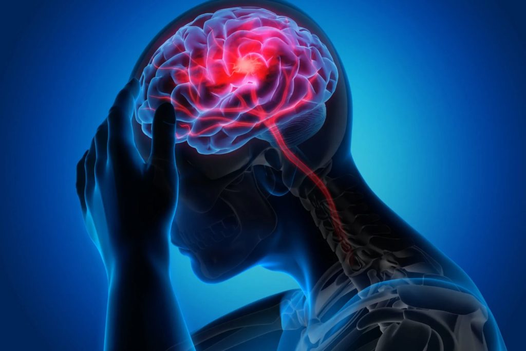
Study shows how aura may lead to migraine headache

- Researchers found that aura altered brain fluid in mice, which flowed directly into part of the peripheral nervous system that detects migraine pain.
- This previously unknown pathway of communication could potentially be targeted to develop new treatments for migraines with aura.
· About a third of people who suffer from migraines experience a phenomenon called aura before the pain sets in. Aura includes visual disturbances and other neurological symptoms. These usually appear within the hour before migraine pain begins.
· Scientists know that a widespread disruption of electrical activity in the brain called cortical spreading depression (CSD) causes aura. But how this disruption might trigger the pain of a migraine has been poorly understood. The sensory neurons that drive migraine pain sit outside the brain. It had previously been thought that the blood-brain barrier lies between the areas of the brain where CSD happens and the neurons that trigger migraine. This could prevent signaling molecules caused by CSD from reaching these neurons.
· A research team led by Dr. Martin Kaag Rasmussen, formerly at NIH and now at the University of Copenhagen, and Dr. Maiken Nedergaard of the University of Rochester has been studying ways that fluids flow through and from the brain. In their new study, funded in part by NIH, they tracked the flow of cerebrospinal fluid from the brain to the peripheral nervous system in mice after CSD. Their results were published on July 5, 2024, in science.
· Using advanced imaging techniques, the researchers observed markers injected into the cerebrospinal fluid flow from the brain rapidly and directly into a peripheral nerve structure called the trigeminal ganglion. Molecules injected into the cerebrospinal fluid were able to activate receptors on cells in the trigeminal ganglion, indicating a direct route of chemical communication.
· Further imaging work found that the membrane that prevents external molecules from entering the trigeminal ganglion was lacking from one end of the ganglion. This created a path for cerebrospinal fluid to flow directly from the brain into the ganglion.
· The team next traced the path of molecules injected directly into the visual cortex. This part of the brain is the most common site of aura. They found that a compound injected into the visual cortex of mice made its way into the trigeminal ganglion in about half an hour. This time delay corresponds to the typical interval between aura and headache onset.
· To better understand the actual substances released by the brain after aura, the researchers induced CSD in mice and then measured changes in proteins found in their cerebrospinal fluid. After CSD, levels of more than 150 proteins changed in cerebrospinal fluid. Twelve of these bound directly to receptors in the ganglion. These included proteins known to be involved in migraine headache, like calcitonin gene-related peptide, or CGRP. It also included other proteins whose potential role in headache need further study.
· “These findings provide us with a host of new targets to suppress sensory nerve activation to prevent and treat migraines and strengthen existing therapies,” Nedergaard says.
· The changes in proteins observed in the cerebrospinal fluid after aura were short lived. It’s therefore likely that other processes play a role in the long duration of migraine headache. More work is needed to uncover these processes and understand how they might be targeted therapeutically.
References: Trigeminal ganglion neurons are directly activated by influx of CSF solutes in a migraine model. Kaag Rasmussen M, Møllgård K, Bork PAR, Weikop P, Esmail T, Drici L, Wewer Albrechtsen NJ, Carlsen JF, Huynh NPT, Ghitani N, Mann M, Goldman SA, Mori Y, Chesler AT, Nedergaard M. Science. 2024 Jul 5;385(6704):80-86. doi: 10.1126/science.adl0544. Epub 2024 Jul 4. PMID: 38963846.
2.Understanding how exercise affects the body
At a Glance
- A study of endurance training in rats found molecular changes throughout the body that could help explain the beneficial effects of exercise on health.
- Large differences were seen between male and female rats, highlighting the need to include both women and men in exercise studies.
· Exercise is one of the most beneficial activities that people can engage in. Regular exercise reduces the risk of heart disease, diabetes, cancer, and other health problems. It can even help people with many mental health conditions feel better.
· But exactly how exercise exerts its positive effects hasn’t been well understood. And different people’s bodies can respond very differently to certain types of exercise, such as aerobic exercise or strength training.
· Understanding how exercise impacts different organs at the molecular level could help health care providers better personalize exercise recommendations. It might also lead to drug therapies that could stimulate some of the beneficial effects of a workout for people who are physically unable to exercise.
· To this end, researchers in the large, NIH-funded Molecular Transducers of Physical Activity Consortium (MoTrPAC) have been studying how endurance exercise and strength training affect both people and animals. The team is examining gene activity, protein alterations, immune cell function, metabolite levels, and numerous other measures of cell and tissue function. The first results, from rat studies of endurance exercise, were published on May 2, 2024, in Nature and several related journals.
· Both male and female rats underwent progressive exercise training on a treadmill over an 8-week period. By the end of training, male rats had increased their aerobic capacity by 18%, and females by 16%. Tissue samples were collected from 18 different organs, plus the blood, during the training period and two days after the final bout of exercise. This let the researchers study the longer-term adaptations of the body to exercise.
· Changes in gene activity, immune cell function, metabolism, and other cellular processes were seen in all the tissues studied, including those not previously known to be affected by exercise. The types of changes differed from tissue to tissue.
· Many of the observed changes hinted at how exercise might protect certain organs against disease. For example, in the small intestines, exercise decreased the activity of certain genes associated with inflammatory bowel disease and reduced signs of inflammation in the gut. In the liver, exercise boosted molecular changes associated with improved tissue health and regeneration.
· Some of the effects differed substantially between male and female rats. For example, in male rats, the eight weeks of endurance training reduced the amount of a type of body fat called subcutaneous white adipose tissue (scWAT). The same amount of exercise didn’t reduce the amount of scWAT in female rats. Instead, endurance exercise caused scWAT in female rats to alter its energy usage in ways that are beneficial to health. These and other results highlight the importance of including both women and men in exercise studies.
· The researchers also compared gene activity changes in the rat studies with those from human samples taken from previous studies and found substantial overlap. They identified thousands of genes tied to human disease that were affected by endurance exercise. These analyses show how the MoTrPAC results from rats can be used to help guide future research in people.
· “This is the first whole-organism map looking at the effects of training in multiple different organs,” says Dr. Steve Carr, a MoTrPAC investigator from the Broad Institute. “The resource produced will be enormously valuable, and has already produced many potentially novel biological insights for further exploration.”
References: Temporal dynamics of the multi-omic response to endurance exercise training. MoTrPAC Study Group; Lead Analysts; MoTrPAC Study Group. Nature. 2024 May;629(8010):174-183. doi: 10.1038/s41586-023-06877-w. Epub 2024 May 1. PMID: 38693412.
Weight-loss surgery yields long-term benefits for type 2 diabetes

Bariatric surgery helped people with type 2 diabetes better control their blood glucose years later compared to medical and lifestyle interventions.
The findings support the use of weight-reduction surgery for treating type 2 diabetes in people with obesity.
· Diabetes affects more than 38 million people nationwide. It occurs when levels of blood sugar, or glucose, are too high. Over time, excess blood glucose can lead to serious health problems, such as heart disease, stroke, nerve damage, and eye disease.
· Some people with type 2 diabetes—the most common type—keep blood glucose in check by making lifestyle changes, including diet and exercise. Medications can also help to control blood glucose. Clinical trials over the past few decades have found that bariatric surgery, or weight-control surgery, can also help control type 2 diabetes. But it had been unclear which of these interventions might have better long-term outcomes.
· To learn more, NIH-supported researchers at four institutions drew on data collected from four previous clinical trials conducted between May 2007 and August 2013. These trials were single-centre studies comparing the effectiveness of bariatric surgeries to medical and lifestyle interventions. The surgeries included sleeve gastrectomy, Roux-en-Y gastric bypass, and adjustable gastric banding. The medical and lifestyle interventions included nutrition counselling, self-monitoring of glucose, and medication to treat diabetes. By pooling data from the four clinical trials, the researchers had a larger, more diverse data set to analyse. Follow-up data was collected 7 to 12 years after the start of the original trials, through July 2022.
· In total, 262 study participants agreed to long-term follow-up. All were between ages 18 and 65. Each had overweight or obesity, as measured by body mass index (BMI). Nearly 70% of participants were women, 31% were Black, and 67% were white. More than half (166) were randomized to receive bariatric surgery. The remaining 96 received diabetes medications plus lifestyle interventions known to be effective for weight loss. Results appeared in the Journal of the American Medical Association on February 27, 2024.
· The researchers found that, seven years after the original intervention, 54% of those in the surgery group had an A1c measurement less than 7%. A1c is a blood test that measures a person’s average blood sugar levels over the previous two or three months. In contrast, only 27% of those in the medical/lifestyle group had similar A1c values.
· In addition, 18% of those in the surgery group no longer had signs or symptoms of diabetes by year seven, compared to 6% in the medical/lifestyle group. The surgery group also had an average weight loss of 20%, compared to 8% in the other group. The differences between groups remained significant at 12 years.
· No differences in major side effects were detected. The surgery group did have a higher number of fractures, anemia, low iron, and gastrointestinal events. These might have been due to greater weight loss and associated nutritional deficiencies. Sleeve gastrectomy and Roux-en-Y gastric bypass were both better than adjustable gastric banding at reducing A1c levels.
· The surgeries appeared to be beneficial even among those with lower BMI scores, between 27 and 34 at study enrollment. That BMI range includes overweight and low-range obesity. Such people had typically been excluded from receiving bariatric surgery for diabetes. But this finding aligns with other recent data that support the use of surgery for some people with a BMI less than 35.
· “These results show that people with overweight or obesity and type 2 diabetes can make long-term improvements in their health and change the trajectory of their diabetes through surgery,” says Dr. Jean Lawrence of NIH’s National Institute of Diabetes and Digestive and Kidney Diseases.
· References: L ong-Term Outcomes of Medical Management vs Bariatric Surgery in Type 2 Diabetes. Courcoulas AP, Patti ME, Hu B, Arterburn DE, Simonson DC, Gourash WF, Jakicic JM, Vernon AH, Beck GJ, Schauer PR, Kashyap SR, Aminian A, Cummings DE, Kirwan JP. JAMA. 2024 Feb 27;331(8):654-664. doi: 10.1001/jama.2024.0318. PMID: 38411644.
Protect Your Child from Dehydration and Heat Illness

As the temperatures rise, it’s crucial to safeguard your child from dehydration and heat-related illnesses. Whether they’re playing outdoors, attending sports events, or simply enjoying summer activities, children are more susceptible to dehydration and heat exhaustion than adults. At Natus Women & Children Hospital, we prioritize your child’s health and well-being, offering expert guidance on preventing and managing these conditions.
Understanding Dehydration and Heat Illness:
Dehydration occurs when the body loses more fluids than it takes in, leading to an imbalance in electrolytes and vital fluids. Heat illness encompasses a range of conditions, including heat exhaustion and heatstroke, which result from prolonged exposure to high temperatures and inadequate hydration.
Signs and Symptoms:
Recognizing the signs of dehydration and heat illness is essential for prompt intervention. Common symptoms include:
· Thirst and dry mouth
· Decreased urine output or dark-coloured urine
· Fatigue and weakness
· Dizziness or light-headedness
· Headache
· Nausea or vomiting
· Muscle cramps
· Rapid heartbeat and breathing
Preventive Measures:
To protect your child from dehydration and heat illness, follow these preventive measures:
· Stay Hydrated: Encourage your child to drink plenty of fluids throughout the day, especially water. Avoid sugary drinks and caffeinated beverages, as they can contribute to dehydration.
· Dress Appropriately: Choose lightweight, loose-fitting clothing in breathable fabrics like cotton to help your child stay cool and comfortable in hot weather.
· Seek Shade: Limit outdoor activities during the hottest part of the day, typically between 10 a.m. and 4 p.m. When outdoors, find shaded areas where your child can rest and cool off.
· Apply Sunscreen: Protect your child’s skin from sunburn by applying sunscreen with SPF 30 or higher before heading outdoors.
· Take Breaks: Encourage regular breaks during physical activities to prevent overheating. Allow your child to rest in a cool, shaded area and offer them water or electrolyte-rich drinks.
· Monitor Activity Levels: Pay attention to your child’s activity level and adjust accordingly. Avoid intense physical exertion during hot weather, and encourage indoor activities when temperatures soar.
· Know When to Seek Help: Be vigilant for signs of dehydration and heat illness, and seek medical attention if your child exhibits severe symptoms such as confusion, fainting, or seizures.
At Natus Women & Children Hospital, our team of experienced paediatricians is dedicated to providing comprehensive care for children of all ages. If you have any concerns about dehydration or heat illness, don’t hesitate to reach out to the best paediatric hospital in Bangalore for expert guidance and support.
By following these tips and staying vigilant, you can protect your child from dehydration and heat-related illnesses, ensuring a safe and enjoyable summer season. Remember, prevention is key, so take proactive steps to keep your child healthy and hydrated in hot weather.
How To Increase Haemoglobin During Pregnancy

During pregnancy, maintaining adequate haemoglobin levels is crucial for both the mother’s and baby’s health. Haemoglobin is responsible for carrying oxygen to the tissues and organs, and low levels can lead to complications such as fatigue, weakness, and even preterm birth. If you’re struggling with low haemoglobin levels during pregnancy, don’t worry! There are several ways to increase haemoglobin naturally and safely. In this blog, we’ll explore some effective tips and advice to boost your haemoglobin levels and ensure a healthy pregnancy journey.
Eat Iron-Rich Foods:
Iron is essential for haemoglobin production, so incorporating iron-rich foods into your diet is key. Include foods such as beans, lentils, tofu, spinach, kale, broccoli, and fortified cereals in your meals. Consuming vitamin C-rich foods like citrus fruits, strawberries, bell peppers, and tomatoes alongside iron-rich foods can enhance iron absorption.
Take Iron Supplements:
In addition to dietary sources, your obstetrician may recommend iron supplements to meet your increased iron needs during pregnancy. Iron supplements help prevent or treat iron deficiency anemia, a common condition characterized by low haemoglobin levels. It’s essential to follow your doctor’s advice regarding the dosage and timing of iron supplements to ensure optimal absorption and minimize side effects like constipation.
Consider Vitamin Supplements:
Apart from iron, other vitamins and minerals also play a role in haemoglobin synthesis. Folate (vitamin B9) and vitamin B12 are essential for red blood cell production and can help increase haemoglobin levels. Your obstetrician may prescribe prenatal vitamins containing these nutrients to support your overall health and well-being during pregnancy.
Stay Hydrated:
Drinking an adequate amount of water is crucial for maintaining blood volume and circulation, which can indirectly impact haemoglobin levels. Aim to drink at least 8-10 glasses of water per day, and consider incorporating hydrating fluids like coconut water, herbal teas, and fruit-infused water into your routine.
Avoid Iron Inhibitors:
Certain substances can interfere with iron absorption and hold back your efforts to increase haemoglobin levels. Limit or avoid consumption of tea, coffee, calcium-rich foods, and high-fibre foods like whole grains and bran cereals close to mealtimes, as they can inhibit iron absorption. Instead, consume these items at least 1-2 hours before or after iron-rich meals.
Practice Healthy Eating Habits:
Maintaining a balanced diet and eating regular, nutritious meals can help support optimal haemoglobin levels during pregnancy. Eat variety of foods like fruits, vegetables, whole grains, lean proteins, and healthy fats. Avoid excessive consumption of processed foods, sugary snacks, and fried foods, which offer little nutritional value.
Monitor Your Haemoglobin Levels:
Regular prenatal check-ups with the best obstetrician in Mysore Road are essential for monitoring your haemoglobin levels and overall health during pregnancy. Your doctor will perform blood tests to assess your haemoglobin levels and provide personalized recommendations based on your specific needs and medical history.
Increasing haemoglobin levels during pregnancy is achievable with the right approach and guidance from the best maternity hospital in Mysore Road. By following a balanced diet, taking supplements as recommended, staying hydrated, and adopting healthy lifestyle habits, you can support your body’s haemoglobin production and ensure a healthy pregnancy journey. Consult the best pregnacy care hospital in Mysore Road for expert care and support throughout your pregnancy.
